
Case
137 With discussion |
Versión
en Español |
|||
CASE 137 (July 2017)
Clinical information
The patient is an 11-year-old girl with terminal chronic kidney disease of unknown cause. Transplantation and nephrectomy of one of the native kidneys is done to try to determine the etiology of her kidney disease.
Clinical history: Birth to term, with low birth weight, without other peripartum alterations. Delayed growth, without alterations in mental or motor development. Polyuria and polydispsia (according to mother's questioning). In urine studies, subnephrotic proteinuria was documented: 26 mg/m2/h. No family history of kidney disease. Studies for viruses and autoimmunity: negative.
Renal ultrasonography: small asymmetric kidneys with poor corticomedullary differentiation, right: 5.1 x 3.8 x 3.3 cm, left: 4.2 x 3.0, x 3.0 cm, with no cysts or dilatation of pyelocaliceal cavities.
See the images.

Figure 1. Fragments of nephrectomy specimen after selection of sections for histology.

Figure 2. H&E, X100.

Figure 3. Methenamine-silver staining, X100.

Figure 4. H&E, X200.

Figure 5. H&E, X400.

Figure 6. H&E, X400.

Figure 7. H&E, X200.

Figure 8. H&E, X400.

Figure 9. Methenamine-silver staining, X400.
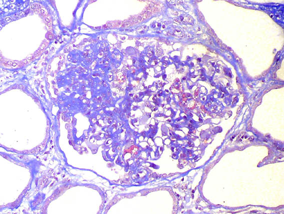
Figure 10. Masson's trichrome staining, X400.

Figure 11. H&E, X400.
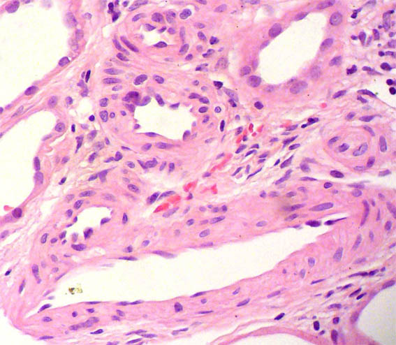
Figure 12. H&E, X400.
Direct immunofluorescence for IgA, IGg, IgM, C3, and C1q: Negative.
What is your diagnosis?