
Case
214 With discussion |
Versión
en Español |
|||
CASE 214 (December 2023)
Clinical information
A 2-year-old boy is evaluated because his mother sees and palpates a bulging in the left abdomen. Ultrasonography demonstrates a multichytic renal mass that replaces approximately 80% of the left kidney. There are no alterations in the right kidney. No other lesions are detected in other organs. The child has normal growth and development. There is no weight loss and the lesion is asymptomatic.
A nephrectomy is performed. Look at the images.
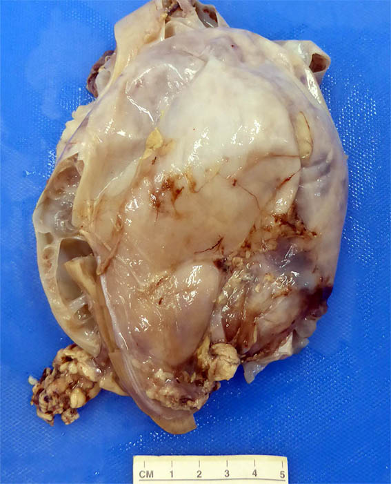
Figure 1. External aspect of the kidney, previously opened.
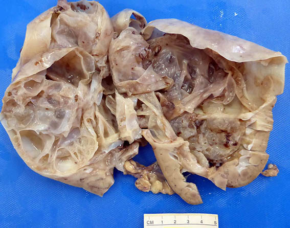
Figure 2. Well-defined multicystic mass, only a remaining portion of renal parenchyma is identified in the lower pole.
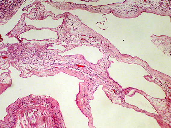
Figure 3. H&E, X100.
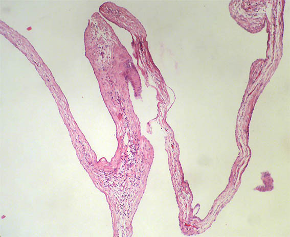
Figure 4. H&E, X100.
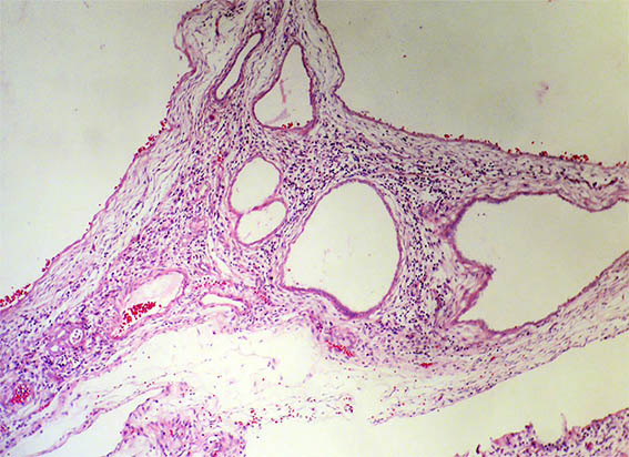
Figure 5. H&E, X100.
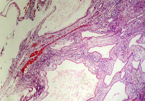
Figure 6. H&E, X100.
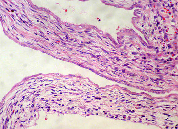
Figure 7. H&E, X200.

Figure 8. H&E, X400.
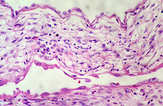
Figure 9. H&E, X400.
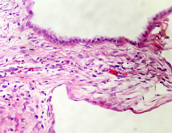
Figure 10. H&E, X400.
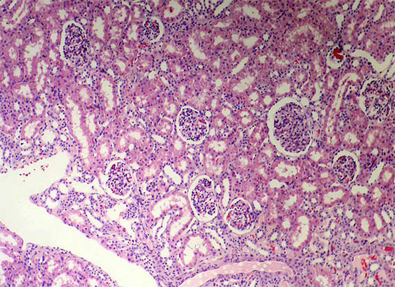
Figure 11. H&E, X100. No alterations were identified in the non-neoplastic parenchyma.
What is your diagnosis?