
Case
213 With discussion |
Versión
en Español |
|||
CASE 213 (November 2023)
Clinical information
A 62-year-old woman is evaluated for erythema on both legs that has been developing for several weeks. There is no personal pathological history. On physical examination, they found systemic arterial hypertension, edema of the lower limbs, purpuric lesions on both legs, and Raynaud's phenomenon. In paraclinical tests, the hemoleukogram is normal; serum creatinine: 2.1 mg/dL, BUN: 39 mg/dL. ANA, anti-DNA, and ANCA: negative. C3: 82 mg/dL (84-180) and C4: 4.1 mg/dL (12-40). HIV and hepatotropic viruses: negative. In urinalysis there is microhematuria, leukocytes: 5/hpf, proteinuria: 4.2 g/24 hours, with mild hypoalbuminemia.
Look at the images.
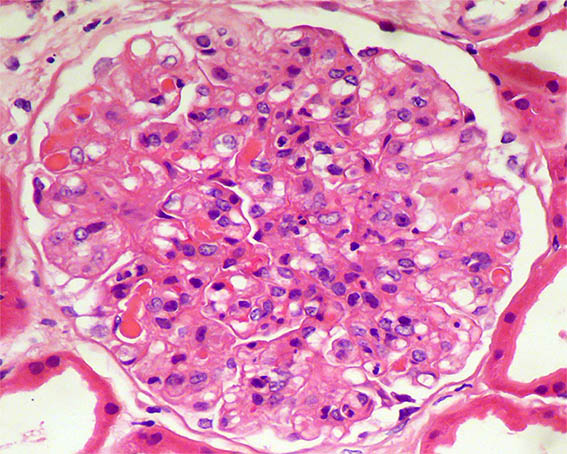
Figure 1. H&E, X400.
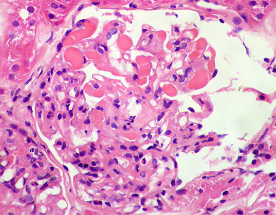
Figure 2. H&E, X400.
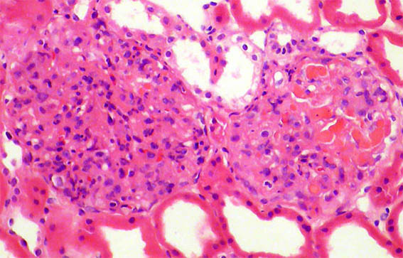
Figure 3. H&E, X200.
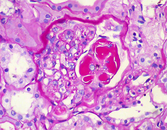
Figure 4. PAS, X400.
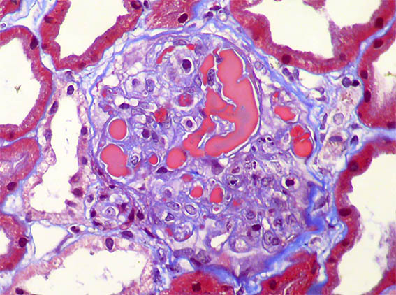
Figure 5. Masson's trichrome stain, X400.
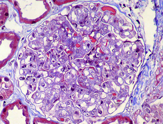
Figure 6. Masson's trichrome stain, X400.
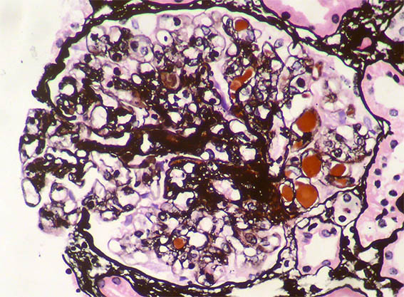
Figure 7. Methenamine-silver stain, X400.
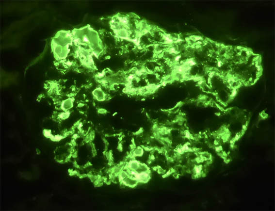
Figure 8. Direct immunofluorescence for IgG, X400.
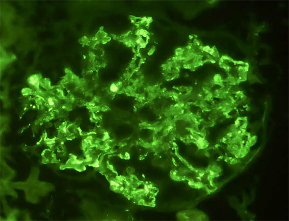
Figure 9. Direct immunofluorescence for kappa light chain, X400.
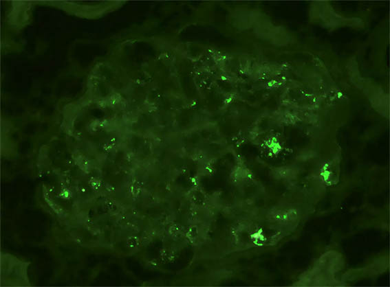
Figure 10. Direct immunofluorescence for lambda light chain, X400.
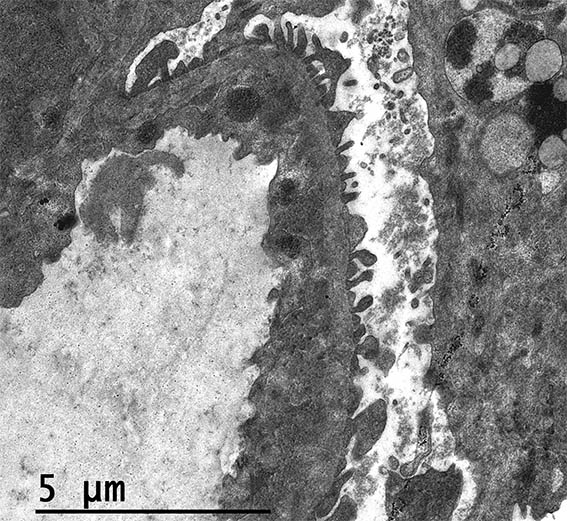
Figure 11. Electron microscopy, original magnification, X2,500. There is endothelial edema and subendothelial electron-dense deposits.
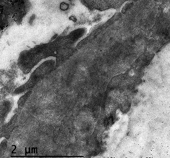
Figure 12. Electron microscopy, original magnification, X4.000. Irregular thickening of the GBM and electron-dense subendothelial deposits.
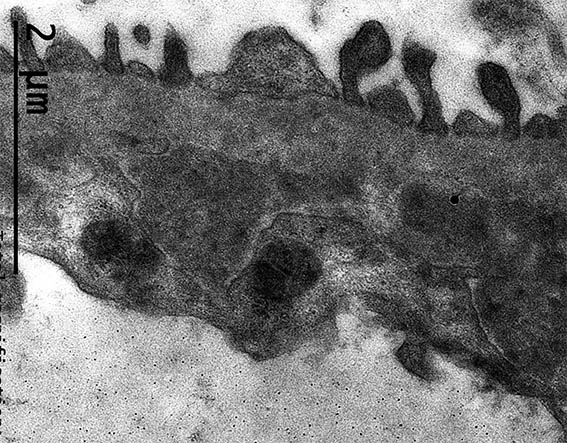
Figure 13. Electron microscopy, original magnification, X4.000. Subendothelial unorganized electron-dense deposits. Endothelium with loss of fenestrations due to edema.
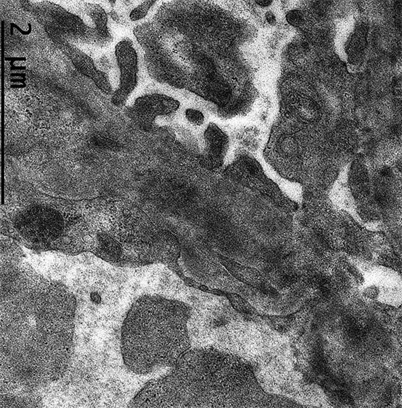
Figure 14. Electron microscopy, original magnification, X4.000.
In immunofluorescence there is also positivity for C3 and, somewhat weaker, for C1q. IgA and IgM: Negative.
What is your diagnosis?