
Case
209 With discussion |
Versión
en Español |
|||
CASE 209 (July 2023)
Clinical information
A 53-year-old patient diagnosed with HIV infection 2 years earlier, on antiretroviral treatment, is evaluated for 2 weeks of edema, with no other symptoms. In laboratory tests, the hemoleukogram is normal. CD4 count: 510 /mm2; viral load: less than 500 copies / mL. Nephrotic syndrome was documented, with proteinuria of 7.5 g/24h, hypoalbuminemia, and dyslipidemia. Serum creatinine: 1.0 mg/dL, BUN: 14.0 mg/dL. Studies for hepatitis virus: negative. ANAs and anti-DNA negative; normal serum complement; Non-reactive VDRL. Serum protein electrophoresis: normal.
Renal biopsy is done, look at the images.
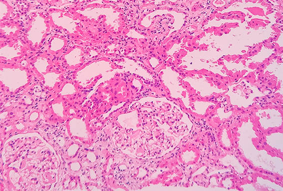
Figure 1. H&E, X200.
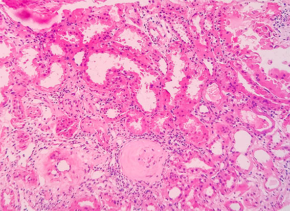
Figure 2. H&E, X200.
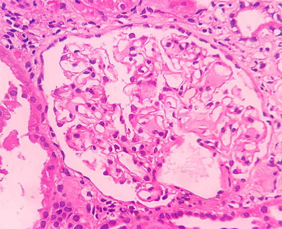
Figure 3. H&E, X400.

Figure 4. H&E, X400.
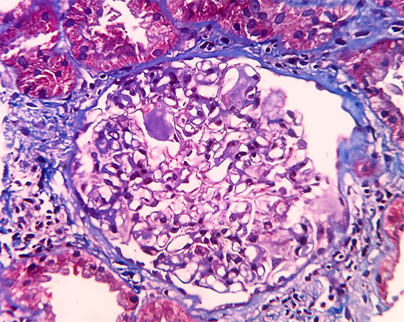
Figure 5. Masson's trichrome stain, X400.
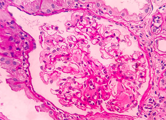
Figure 6. PAS, X400.
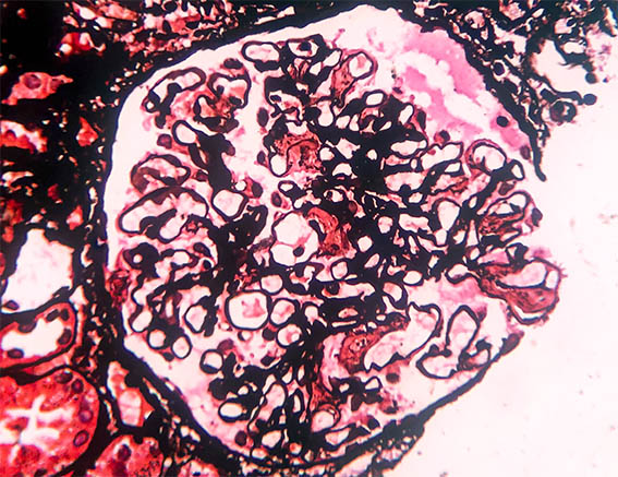
Figure 7. Methenamine-silver stain, X400.
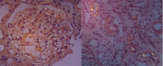
Figure 8. Congo red staining seen with polarized light, X400. It was positive, although the digital images do not allow a quality view of the green birefringence.
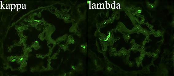
Figure 9. Immunofluorescence for kappa and lambda, X400. Both negative.
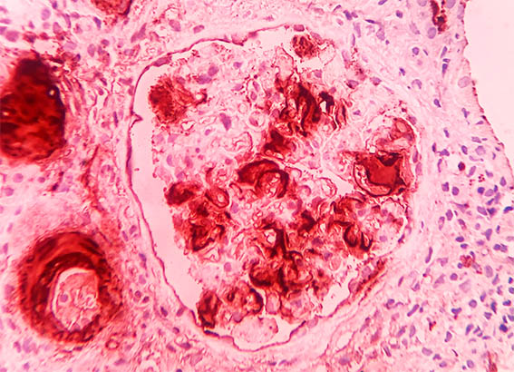
Figure 10. Immunohistochemistry for AA amyloid, X400.
Direct immunofluorescence for IgA, IgG, IgM, C3 and C1q: Negative.
What is your diagnosis?
What is the relationship with HIV infection?