
Case
208 With discussion |
Versión
en Español |
|||
CASE 208 (June 2023)
Clinical information
A 43-year-old man with end-stage renal failure of unknown cause, presumably glomerulopathy, was transplanted from a cadaveric donor. Adequate renal function during the first 2 years, with creatinine between 0.8 and 1.0 mg/dL. Immunosuppression scheme: prednisone, tacrolimus and mycophenolate, with adequate adherence and serum levels of tacrolimus within the expected therapeutic range. In a periodic control, the third year after the transplant, serum creatinine was elevated: 1.4 mg/dL, BUN: 27 mg/dL. Urinalysis with proteinuria: 300 mg/dL, microhematuria: 15-20 erythrocytes/hpf, some dysmorphic, leukocytes: 6-8/hpf; 24-hour urine protein: 2.6 g/24h. Infectious disease studies negative, normal complement. Resistance indices in graft vessels: normal.
A biopsy of the renal graft is performed, observe the images.
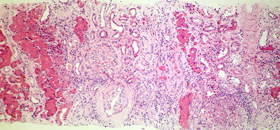
Figure 1. H&E, X100.
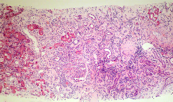
Figure 2. H&E, X100.
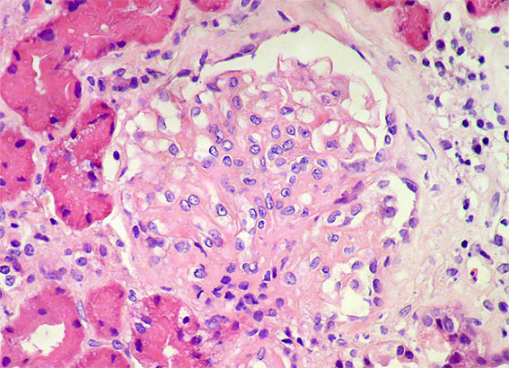
Figure 3. H&E, X400. Segmental sclerosing lesion.
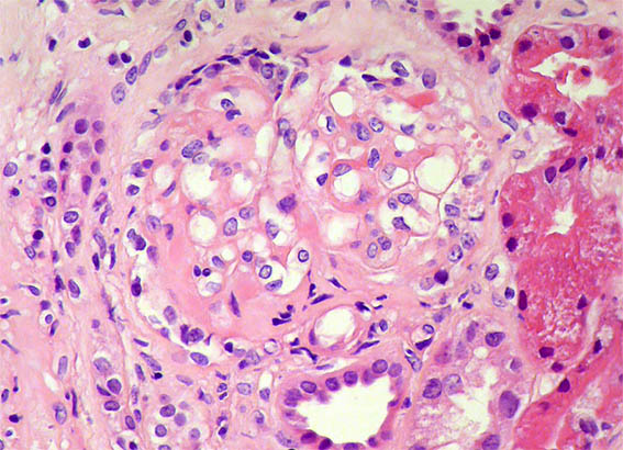
Figure 4. H&E, X400. Segmental sclerosing lesion.
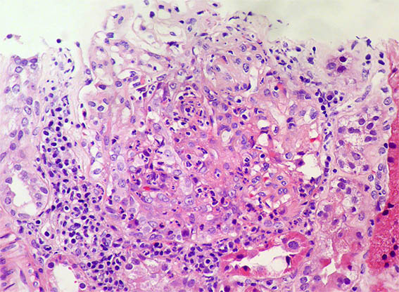
Figure 5. H&E, X400. Endocapillary hypercellularity (exudative) and a possible cresent.
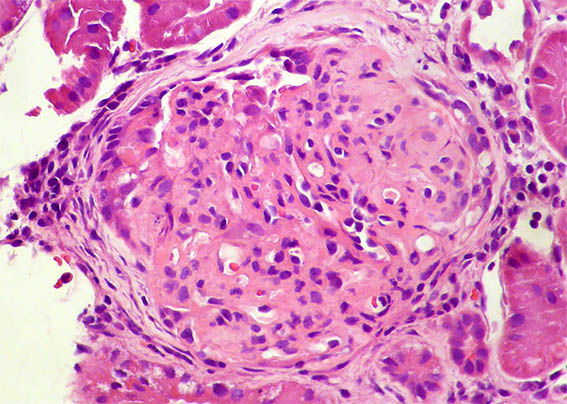
Figure 6. H&E, X400. Segmental sclerosis. Fragment fixed in glutaraldehyde.
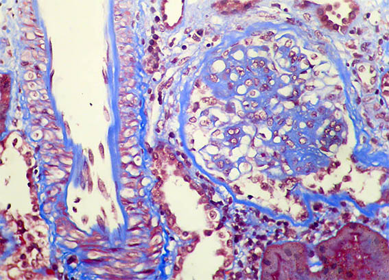
Figure 7. Masson's trichrome stain, X400. Mesangial expansión; normal artery.
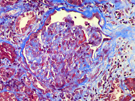
Figure 8. Masson's trichrome stain, X400.
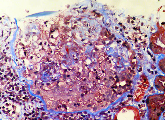
Figure 9. Masson's trichrome stain, X400. A crescent.
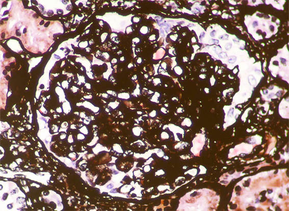
Figure 10. Methenamine-silver stain, X400.
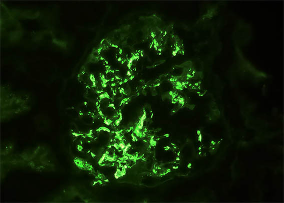
Figure 11. Immunofluorescence for IgA, X400. Mesangial and subendothelial positivity.
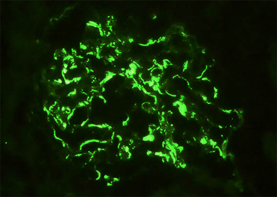
Figure 12. Immunofluorescence for IgM, X400. Something similar to IgA.
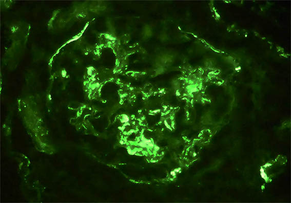
Figure 13. Immunofluorescence for C3, X400.
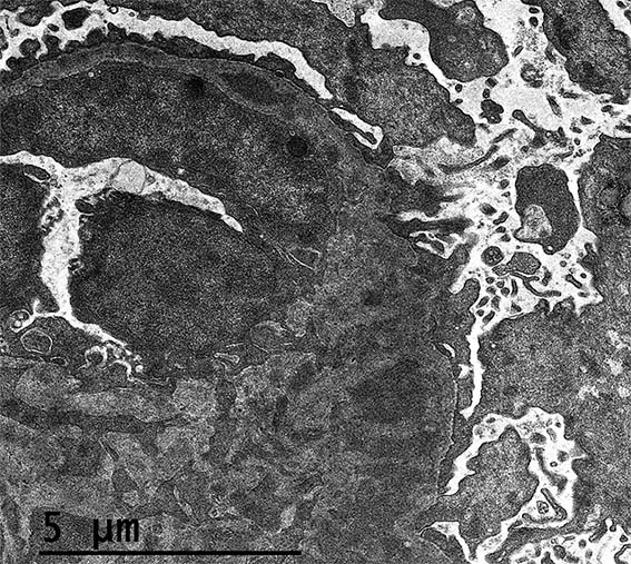
Figure 14. Electron microscopy, original magnification, X2.100. Mesangial and subendothelial electron-dense deposits; note endothelial edema.
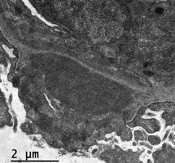
Figure 15. Electron microscopy, original magnification, X2.550. A subepithelial deposit.
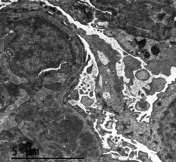
Figure 16. Electron microscopy, original magnification, X2.100. Mesangial electron-dense deposits and a capillary lumen almost obstructed by a cell.
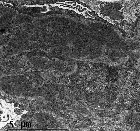
Figure 17. Electron microscopy, original magnification, X2.550. Mesangial electron-dense deposits.
Direct immunofluorescence for IgG and C1q: Negative; kappa y lambda: similar staining to that of IgA and IgM. C4d: Negative.
What is your diagnosis?