
Case
204 With discussion |
Versión
en Español |
|||
CASE 204 (February 2023)
Clinical information
A 65-year-old woman with a history of insulin-requiring diabetes mellitus presents generalized edema of 10 days' evolution. Nephrotic syndrome is documented, with proteinuria of 15 g/24 hours; serum albumin: 2.1 g/dL. Two months earlier, in a follow-up exam, she had proteinuria of 1.2 g/24 hours. The patient also has diabetic retinopathy. In the urinalysis there is microhematuria. Other paraclinical findings: normal hemoleukogram; HbA1c of 10%; serum creatinine: 1.3 mg/dL, BUN: 21 mg/dL. ANAs: 1:160, speckled, anti-DNA: 1:10; C3: 88 (90-180), C4: 11 (12-40). There are no mucocutaneous or joint alterations on physical examination.
Given the rapid increase in proteinuria and the presence of microhematuria, a renal biopsy was performed.
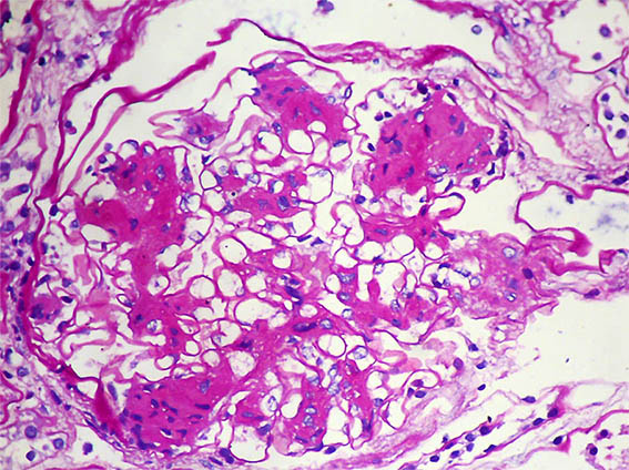
Figure 1. PAS, X400. Mesangial nodules.
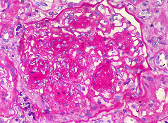
Figure 2. PAS, X400.
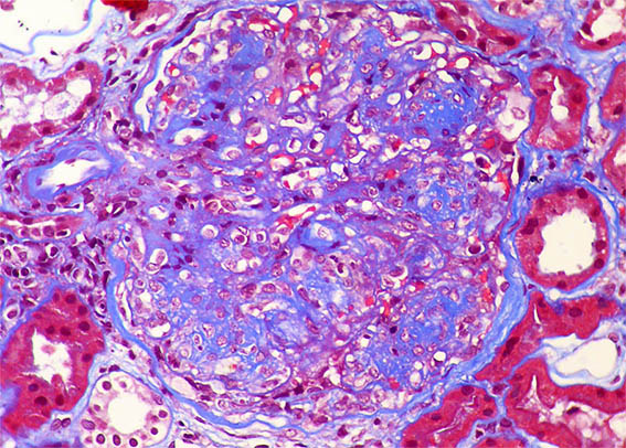
Figure 3. Masson's trichrome stain, X400.
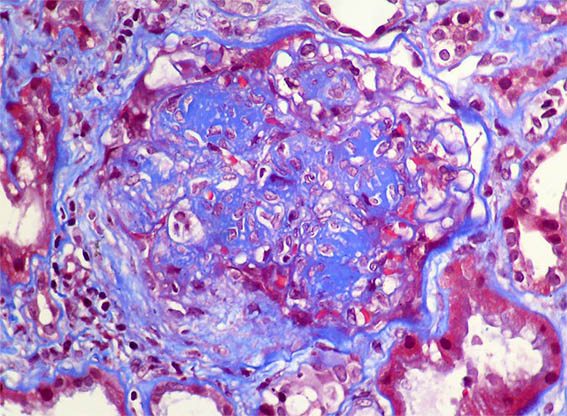
Figure 4. Masson's trichrome stain, X400.
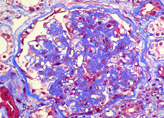
Figure 5. Masson's trichrome stain, X400.
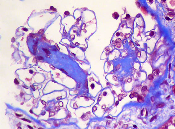
Figure 6. Masson's trichrome stain, X400.
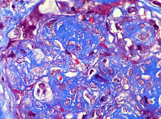
Figure 7. Masson's trichrome stain, X1.000.
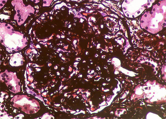
Figure 8. Methenamine-silver stain, X400.
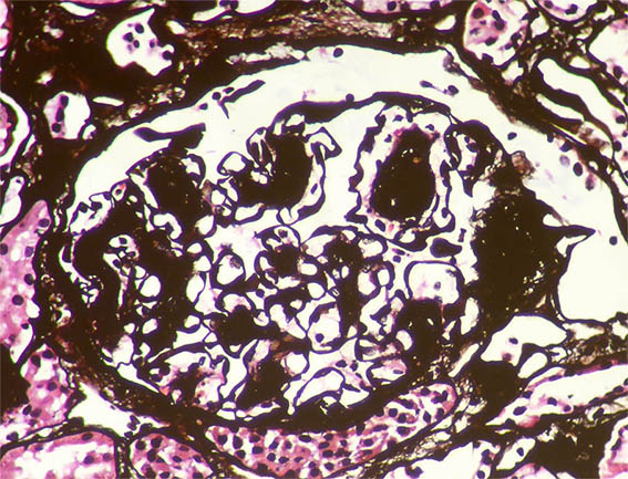
Figure 9. Methenamine-silver stain, X400.
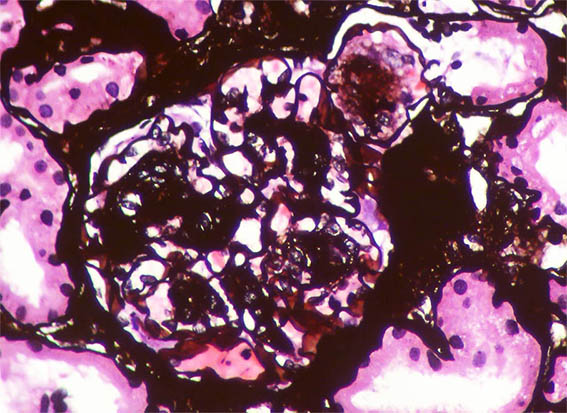
Figure 10. Methenamine-silver stain, X400. Note the nodule on the upper right, surrounded by basement membrane (possibly microaneurysm with organizing thrombus).
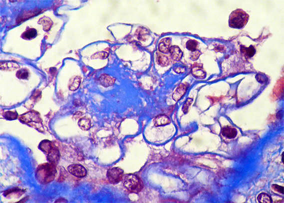
Figure 11. Masson's trichrome stain, X1.000. Some subtle subepithelial fuschinophilic granules.
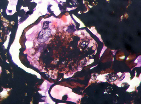
Figure 12. Methenamine-silver stain, X1.000. The same nodule of figure 10; note "holes" in the basement membrane, on the left side.
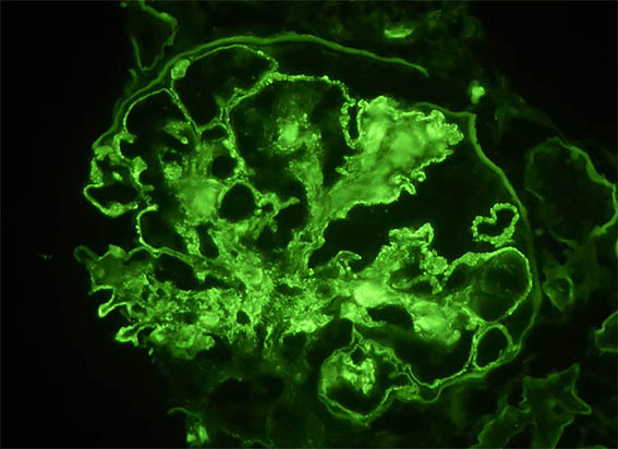
Figure 13. Immunofluorescence for IgG, X400. Subepithelial granular parietal positivity. There is also smesangial positivity.
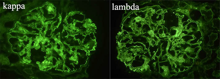
Figure 14. There is positivity for both light chains, with similar intensity (there is no chains restriction), X400.
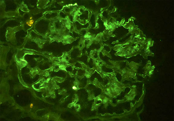
Figure 15. Immunofluorescence for C3, X400. Segmental, weak, irregular positivity.
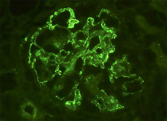
Figure 16. Immunofluorescence for C1q, X400. Subepithelial granular parietal positivity.
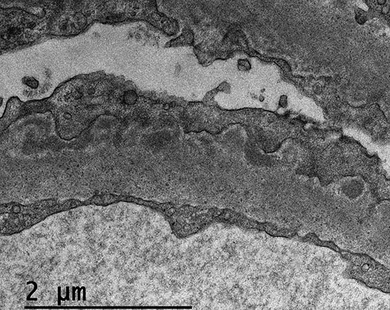
Figure 17. Electron microscopy, original magnification, X4.000. Note electron-dense subepithelial deposits.
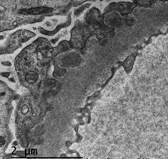
Figure 18. Electron microscopy, original magnification, X4.000. Also here note electron-dense subepithelial deposits.
Direct immunofluorescence for IgA: Negative; IgM: Focal and segmental positivity in hyaline lesions (nonspecific entrapment).
What is your diagnosis?