
Case
202 With discussion |
Versión
en Español |
|||
CASE 202 (December 2022)
Clinical information
The patient is a 23-year-old woman with a history of nephrotic syndrome associated with a genetic alteration (pathogenic mutation, nonsense, in the NUP160 gene, which encodes one of the protein components of the nuclear pore complex nucleoporin 160 kD (Nup160) [Zhao F, et al. Mutations in NUP160 Are Implicated in Steroid-Resistant Nephrotic Syndrome. J Am Soc Nephrol. 2019;30:840-853 [PubMed link]), with biopsy that showed morphology of focal and segmental glomerulosclerosis, she reached terminal renal failure at the age of 21 years. After 2 years on dialysis, she is transplanted from a cadaveric donor. Prior to transplant she was anuric.
Twelve days after the transplant, proteinuria of 12 grams/24h was documented, with edema, dyslipidemia and hypoalbuminemia; serum creatinine is 1.0 mg/dL.
Renal graft biopsy is performed, observe the images:

Figure 1. H&E, X100. No rejection.
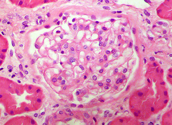
Figure 2. H&E, X400. Glomerulus with normal characteristics.
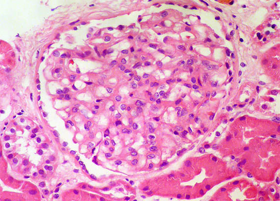
Figure 3. H&E, X400. Perhaps some nonspecific segmental increase in mesangial cellularity.
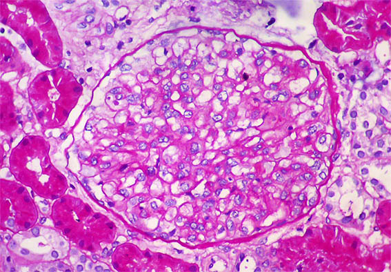
Figure 4. PAS, X400. Without alterations. There are no sclerosing or hyaline lesions in any of the 22 glomeruli in the biopsy.
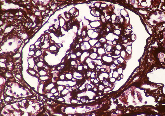
Figure 5. Methenamine-silver stain, X400. Normal capillary walls.
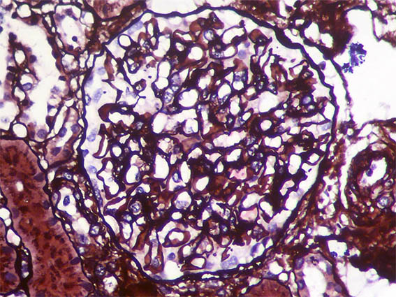
Figure 6. Methenamine-silver stain, X400.
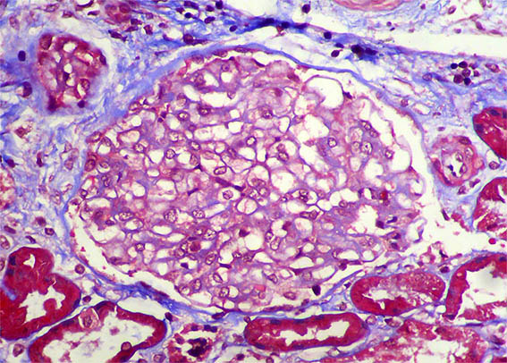
Figure 7. Masson's trichrome stain, X400.
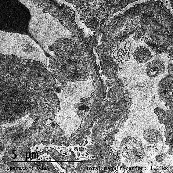
Figure 8. EM, original magnification, X2,100.
![]()
Figure 9. EM, original magnification, X2,100.
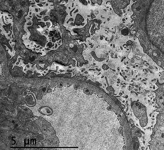
Figure 10. EM, original magnification, X2.100.
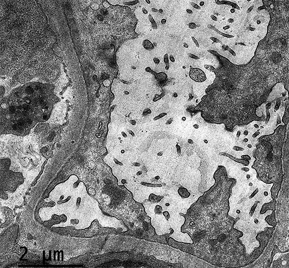
Figure 11. EM, original magnification, X3,000.
![]()
Figure 12. EM, original magnification, X3,000.
Direct immunofluorescence for IgA, IgG, IgM, C3, and C1q: Negative.
What is your diagnosis?